Anesthesia for minimally invasive, video-assisted or robotic-assisted thoracoscopic surgery (VATS), is similar to anesthesia for open thoracic cases in many respects. However, achieving lung isolation quickly and completely is even more important, since even a slightly inflated lung may obstruct the surgeon’s view. Procedures that are amenable to VATS include but are not limited to:
Mediastinoscopy
Wedge resection or lung biopsy
Lobectomy or segmentectomy, including bronchial sleeve resection
Lymph node dissection
Pleurodesis, mechanical or talc, for pleural effusion or spontaneous pneumothorax
Decortication, including evacuation of empyema or hemothorax
Thymectomy
Lung volume reduction for severe emphysema
The intrathoracic portion of esophagectomy
Thoracic sympathectomy for hyperhidrosis or treatment of refractory ventricular tachycardia
Nearly any patient may be a candidate regardless of extremes of age or pulmonary disease.
Procedures usually requiring open thoracotomy include pneumonectomy, intrathoracic tracheal resection, and chest wall resection.
The advantages of VATS include decreased hospital length of stay, decreased morbidity, and less postoperative pain.
The keys to anesthesia success include:
Availability of video laryngoscopy to facilitate intubation;
Facility with placing both right and left endobronchial tubes;
Skill with fiberoptic bronchoscopy;
Having a dedicated team of personnel with expertise in thoracic anesthesiology.
Primary anesthetic considerations include optimal double-lumen tube placement with fiberoptic guidance[1], management of one-lung ventilation to avoid hypoxia and unnecessary hyperoxia[2], and use of protective ventilation strategies to avoid injury to the ventilated lung.[3][4][5] Acute lung injury following thoracic surgery may lead to the development of ARDS with potentially lethal outcomes.[6][7][8][9]
Patients may present for VATS in sound health or with substantial disease burden.
A young, otherwise healthy patient who presents for thoracic sympathectomy for hyperhidrosis, or bleb resection with mechanical pleurodesis for recurrent spontaneous pneumothorax, may require no preoperative testing.
At the opposite extreme, patients may present with COPD, decreased lung function on the affected side, pleural or pericardial effusion, anemia, poor nutritional status, and effects of neoadjuvant chemotherapy and/or radiation. Whenever possible, prehabilitation in preparation for surgery should be considered to correct anemia, improve nutritional status, and improve functional capacity.
Patient evaluation
System
Considerations
Cardiovascular
Evidence of ventricular or valvular dysfunction, pulmonary hypertension
Respiratory
History of COPD, asthma, pleural effusion, pulmonary fibrosis, sarcoidosis, other pulmonary pathology
Neurologic
Peripheral neuropathy due to chemotherapy; evidence of myasthenia or paraneoplastic syndrome; assess appropriateness for epidural analgesia
Gastrointestinal
Evidence of carcinoid syndrome in cases of lung or endobronchial carcinoid tumor
Hematologic
Hypercoagulability due to underlying malignancy
Labs and studies
Routine preoperative laboratory, EKG, and radiographic studies as appropriate for patient age and institutional guidelines
Consider pulmonary function testing
Consider echocardiography, stress testing
Consider ventilation/perfusion (v/q) scan before major lung resection
Operating room setup
Double-lumen endotracheal tube (DLT) or bronchial blocker as appropriate
Fiberoptic bronchoscope
Video laryngoscope -- may facilitate DLT insertion
Consider arterial line setup and transducer
Patient preparation and premedication
Patients should fast per institutional guidelines. Premedication is at the choice of the anesthesiologist depending on the patient's age and other relevant factors.
Regional and neuraxial techniques
Thoracic epidural analgesia may be considered especially if bilateral VATS is undertaken or if the surgeon thinks there is a high likelihood that conversion to open thoracotomy may be necessary.
The epidural catheter should be inserted at a high enough level that the patient will not have any lumbar motor block and can safely ambulate. Insertion prior to surgery offers the advantage of beginning continuous epidural infusion before the patient emerges from anesthesia. Some anesthesiologists prefer to utilize the epidural infusion along with general anesthesia for the entire case. Others prefer to activate the epidural catheter only near the end of surgery once blood loss is over in order to avoid intraoperative hypotension. There is no conclusive evidence that either approach is superior.
If the patient has a contraindication to epidural catheter placement, other regional techniques may be considered for postoperative pain relief: lumbar spinal opioid analgesia, erector spinae plane block[10], intercostal or paravertebral blocks.
Intraoperative management
The goals of intraoperative management include:
Optimal placement of DLT for effective one-lung ventilation
Avoidance of hyperoxia which may be an exacerbating factor in acute lung injury
Maintaining satisfactory peak airway pressure (preferably less than 25-30 cmH20) and plateau pressure during one-lung ventilation; permissive hypercapnia may be necessary to avoid barotrauma to the ventilated lung
Avoiding excessive administration of crystalloid which been associated with acute lung injury
Monitoring and access
Standard monitors suffice for induction for many patients. Patients presenting for less invasive procedures may not require any more than standard monitoring for the duration of the case; examples might include thoracic sympathectomy for hyperhidrosis, limited lung biopsy for tissue diagnosis, talc pleurodesis for palliation of recurrent pleural effusion, or placement of PleurX catheter.
Arterial monitoring is commonly performed for lung resection, esophagectomy, thymectomy, and other major operative procedures. It enables continuous observation of blood pressure, and arterial blood gas measurement if indicated. Whether the arterial catheter should be inserted prior to or after anesthesia induction is a matter of anesthesiologist preference.
As the administration of fluids is best kept to a modest amount for lung resection, more than one well-functioning peripheral IV catheter may not be necessary. This again is a matter of individual judgment and preference.
Central venous pressure or flow parameter monitoring (e.g. FloTrac, Edwards Lifesciences) may be considered for patients undergoing procedures where substantial blood loss and/or fluid requirements are likely, such as esophagectomy or pleurectomy/decortication. A central venous line may be indicated in any procedure if peripheral veins are inadequate.
Induction and airway management
Induction medications and muscle relaxant choice per anesthesiologist preference. Ketamine may be a useful adjunct, especially for patients with chronic pain or a history of preoperative opioid use.
DLT (typically 35-39 Fr) is inserted and position confirmed with fiberoptic bronchoscopy. Selection of the DLT size is dependent on patient height and weight. A 37 Fr DLT is suitable for most average-size female patients, and 37-39 Fr for most male patients. DLT smaller than 35 Fr does not easily accommodate a fiberoptic bronchoscope.
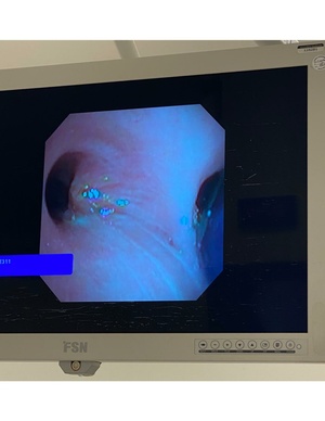
Figure 1: Malignant mucoepidermoid tumor in L mainstem bronchus near the main carina, with R DLT in positionThe selection of right or left DLT is dependent on the situation and on user experience. The only absolute indication for a R DLT is the planned sleeve resection of a L mainstem tumor (Fig. 1).